Treatments & Services Archives - Page 2 of 4 - Michigan Head & Spine Institute Blog

MHSI_web-1319103349-1200x683.jpg


Dr. Fredrick Junn
Fredrick S. Junn, M.D.
Neurosurgeon

While most people have heard of Parkinson’s disease, which affects nearly 1 million people in the United States, fewer of us are aware of its symptoms, risk factors and treatment.

Knowing about Parkinson’s Disease, including early signs to watch for, can help you and your loved ones catch the disease earlier, understand its signs, and learn how to manage it.

What Is Parkinson’s Disease?

Parkinson’s disease is a disorder of the central nervous system that affects movement, specifically how your brain communications with the rest of your body to create and execute movement.

With Parkinson’s disease, damage to nerve cells in the brain lead to reduced levels of dopamine, which plays a role in movement. When dopamine decreases, the brain and the body can’t communicate properly, leading to symptoms of Parkinson’s disease.

Parkinson’s disease is a progressive neurological condition, meaning it starts slowly and gets worse over time. It typically starts in one portion of the body, such as the arm or leg, and eventually crosses over to affect the other side of the body as well. Early- to mid-stage symptoms of Parkinson’s disease include:

  • Tremors/shaking in fingers, thumb, hand or chin
  • Stiffness
  • Constipation
  • Trouble sleeping
  • Smaller handwriting (micrographia)
  • Speaking in a lower or softer voice (hypophonia)
  • Dizziness
  • Slowed movement (bradykinesia)
  • Loss of smell
  • Stooped posture

Very recently, new research has identified two more early signs of Parkinson’s disease: hearing loss and epilepsy.

Since many of these symptoms could be caused by other health conditions, it’s important to talk with your healthcare provider to potentially rule out Parkinson’s disease if you’re experiencing them.

As Parkinson’s disease progresses, so do the symptoms. Some that may show up later in the course of the disease include:

  • Tremors and stiffness on both sides of the body
  • Walking problems
  • Loss of balance
  • Shuffling gait
  • Markedly stooped posture

Parkinson’s disease can also affect a person’s mental health, and is linked to conditions such as depression, anxiety and dementia.

Risk Factors for Parkinson’s Disease

Although scientists haven’t been able to pinpoint what causes Parkinson’s disease, they have identified some risk factors. These include:

  • Genetics
    When people develop Parkinson’s disease at a young age (in their 20s or 30s), the cause is often genetic.
  • Advanced age
    The average age of onset for Parkinson’s disease is around 60 years old.
  • Gender
    Parkinson’s disease is more common in men than women.
  • Environmental triggers
    Exposure to pesticides/herbicides, heavy metals and other toxic chemicals has been shown to increase the risk for developing Parkinson’s disease. A street drug called “synthetic heroin” is also associated with Parkinson’s disease.

Treatments and Management for Parkinson’s Disease

There is no cure for Parkinson’s disease, but it can be managed and even slowed, depending how early the disease is diagnosed. Treating Parkinson’s disease typically involves increasing the amount of dopamine in the brain. This can be done with medications, including some types of antidepressants; newer anti-Parkinsonian medications are designed to provide a steady level of dopamine so patients don’t experience the crash associated with earlier treatments.

In addition to medication, deep brain stimulation (DBS) can be helpful for managing Parkinson’s disease symptoms. DBS is a surgical procedure where electrodes are implanted that deliver tiny electrical impulses to the parts of the brain that control movement. A major advantage of DBS is that it levels off dopamine levels without the side effects associated with some Parkinson’s disease medications.

Keeping the body healthy and moving are also key to controlling Parkinson’s disease symptoms, and many people find physical therapy to be helpful for maintaining movement.

If you think you may be experiencing Parkinson’s disease symptoms, or if you have a family history of the disease, you may want to ask your healthcare provider for a neurological referral. A neurologist can provide a diagnosis and start you down the path of managing your Parkinson’s disease.


If you or a loved one is seeking treatment for a condition of the brain, or to schedule an appointment with Dr. Junn or any of our MHSI experts, visit www.mhsi.com/appointments or call 248-784-3667.

MHSI-1248544072_aneurysm-1200x792.jpg


Dr. John Whapham
John Whapham, M.D., MS, FSNIS, FAAN
Neurosurgeon

Surgery has always been an option for certain types of intracranial cerebral aneurysms, but in recent years, minimally invasive procedures have greatly reduced both physical trauma as well as recovery times.

Cerebral aneurysms occur when part of a blood vessel wall weakens, allowing it to bulge, widen and potentially burst or rupture, which can be life-threatening.

Aneurysms can exist for years without detection. Because of this, there are often no symptoms until an aneurysm ruptures. Once that happens, symptoms may develop suddenly and violently, depending on the location of the aneurysm.

Usually, symptoms come on quickly and are unlike anything you might have experienced before. If you don’t often get headaches but suddenly experience an intense migraine, a ruptured brain aneurysm could be the culprit.

Call 911 and seek immediate medical attention if you or someone you know is having aneurysm signs and symptoms including:

  • Severe headache
  • worst headache of life
  • Stiffness or swelling in the neck
  • loss of consciousness
  • speech difficulty
  • focal motor or sensory symptoms weakness or numbness
  • Dizziness
  • Vision changes
  • Confusion
  • Hoarseness
  • Difficulty swallowing
  • Nausea and vomiting
  • Shock (low blood pressure, rapid heart rate, clammy skin, decreased awareness)

Sometimes an unruptured aneurysm is found when performing unrelated diagnostic testing such as MRIs or CAT scans.

Minimally Invasive Aneurysm Surgery Saves Lives

Aneurysm treatment has greatly improved in the last 25 to 30 years. In the past, major surgery, such as open craniotomy, was commonplace. In this procedure, a small metal clip is placed across the base of the aneurysm bulge that seals off blood flow to the ballooned blood vessel.

However, modern advances have made this major procedure less common.

These days, minimally invasive procedures such as coiling and stenting are the norm. Both divert the flow of blood away from the aneurysm making it less likely to rupture and are performed without opening the skull.

  • Coiling, also called endovascular embolization, uses a catheter passed through the groin up into the artery containing the aneurysm where tiny platinum coils are released. The coils induce clotting of the aneurysm and that clotting prevents blood from entering the aneurysm.
  • Stenting is when a catheter is used to place a stent (a soft, flexible mesh tube) into the blood vessel where an aneurysm has formed. The stent prevents blood from entering the aneurysm. In time, new cells grow on the stent, sealing the aneurysm and healing the vessel.

These minimally invasive procedures allow for a much shorter recovery period, and it is possible to return to daily living as early as one day after hospital discharge.

In fact, some patients with an unruptured aneurysm check in at the hospital, undergo a procedure that is finished in a couple of hours, stay overnight for observation and are discharged the next morning with only a small bandage covering the spot in the groin or wrist where the needle access was made.

Quick Treatment Is Key

It’s crucial to contact first responders at the first sign of an intense, sudden headache since that may indicate a ruptured aneurysm.

If you are diagnosed with an unruptured aneurysm, call us right away to make an appointment.

Depending on the aneurysm’s size and location, you may require regular checkups from a physician trained in aneurysm treatment and surgery. A treatment plan, including lowering your blood pressure, quitting smoking and medical intervention, can help you reduce the risk of rupture.


If you or a loved one is seeking treatment for a condition of the brain, or to schedule an appointment with Dr. Whapham or any of our MHSI experts, visit www.mhsi.com/appointments or call 248-784-3667.

MHSI-Telehealth-1286810754-1200x800.jpg

Dr. Richard D. Fessler
Richard D. Fessler , M.D. Neurosurgeon

In 2013 Dr. Richard Fessler predicted the use of telehealth would become the norm as technology was in place to create visits for patients via a smart phone. Fast forward to 2020, Dr. Fessler saw his prediction become reality. The pandemic of a lifetime gave the entire healthcare industry a push into telehealth. Insurance companies expanded coverage for this type of visit with a push from Center for Medicare Service or CMS to cover costs.

“At MHSI we were ready to offer our patients this option when COVID caused concerns about safety for our patients and staff.  With the insurance barrier removed, it brought reassurance we could deliver the telehealth option and patients didn’t have to be concerned about unnecessary out-of-pocket-costs,” said Dr. Fessler. “We are thankful that our patients accepted the technology and the new way we are able to provide care.”

For example, when Tony couldn’t take his back pain any longer, he met with Neurosurgeon Dr. Ratnesh Mehra virtually and had trust that his surgical experience would be equally the same and relieve his back pain. Dr. Mehra recalls the day they met online, “Tony was able to effectively communicate with me about his pain and what he was feeling. At that point we decided that surgery for Tony was the right course of treatment and I met Tony in pre-op in person for the first time.” Tony says, “One week after surgery I was walking…dancing.”


Watch how intense pain stopped Tony in his tracks!:


To schedule a telehealth appointment with an MHSI neurosurgeon, call 248-784-3667 or visit MHSI.us.

Nasal-Endoscopy_Steven_thumb-1200x772.jpg

Every year neurosurgeons advance the procedures performed to treat brain and spine conditions. Just a few short years ago minimally invasive surgery of any type was unheard of. Yet today, an incision of less than 2 inches allows a neurosurgeon to fix or replace a disc in the neck or spine.

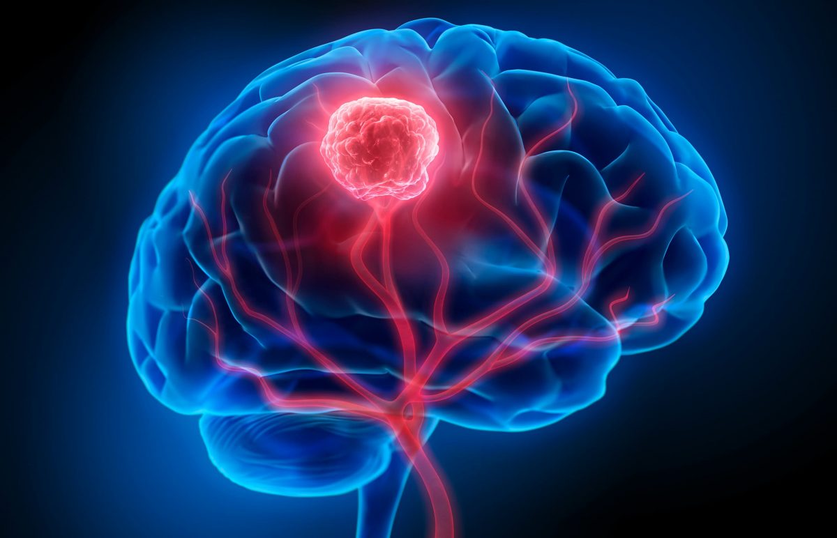
Minimally invasive nasal endoscopy is yet another advancement to treat tumors of the brain. This approach allows the neurosurgeon to access the brain to remove a tumor without performing a craniotomy, which makes a “window” in the bone to allow access to the brain.

Dr. Jeffrey T. Jacob
Jeffrey T. Jacob, M.D. Neurosurgeon

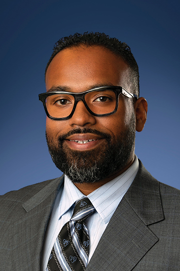
Deciding between a craniotomy or using the nasal cavity to reach the brain, depends on what is best for the patient. “The goal is to be able to reach the tumor and remove all of it,” says Jeffrey Jacob, M.D. “Often, we are able to do that going through the nose with an endoscope, with a very small camera device.”

There are many types of brain tumors that can be treated with this approach. Brain tumors that are removed using the nasal endoscopic approach can be benign or malignant, and typically sit at the base of the skull or under the brain. This procedure removes the tumor and the blood supply to the tumor.

To perform a nasal endoscopy, Dr. Jacob partners with Adam Folbe, M.D., a rhinologist and endoscopic skull base surgeon at Michigan Sinus and Skull Base Center. Dr. Folbe goes through the nose and opens the window to the brain. He says, “There is no cutting of skin, muscle or fat resulting in no scars. Using this approach maximizes retrieval of the tumor with lesser disruption to the brain compared to the craniotomy approach.”

Patients who experience this approach have no signs of surgery on the outside of their body, because the nasal cavity is repaired with the patient’s own tissue, without stitches or sutures. During the post-surgical healing, patients are restricted from sneezing, blowing their nose, lifting and bending over for about three weeks.

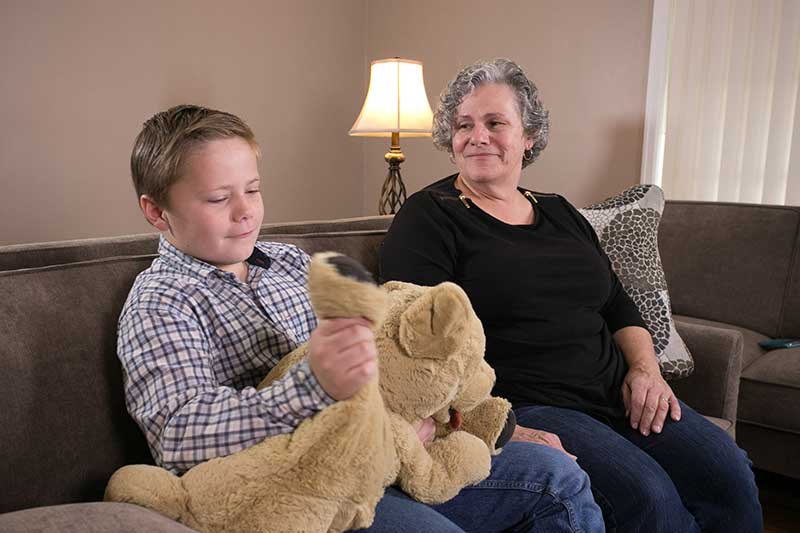
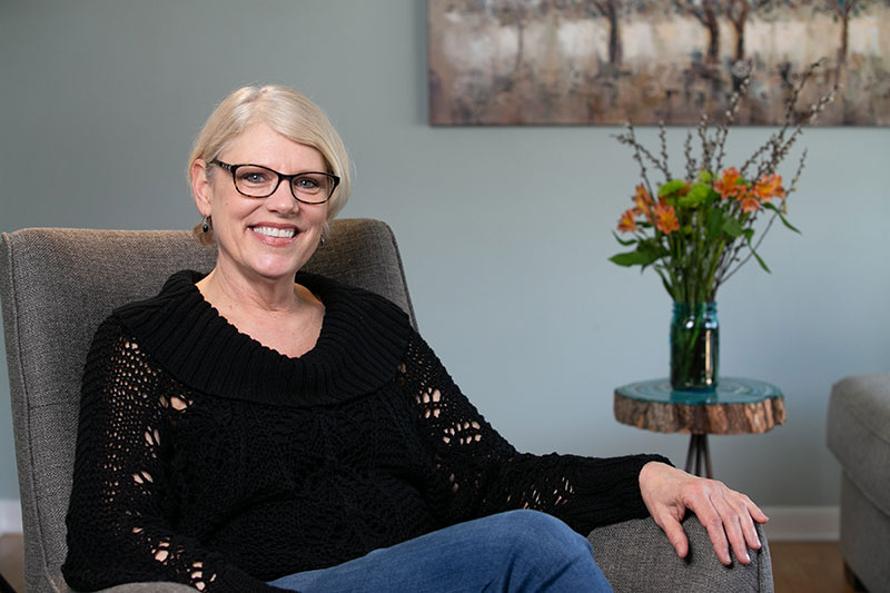
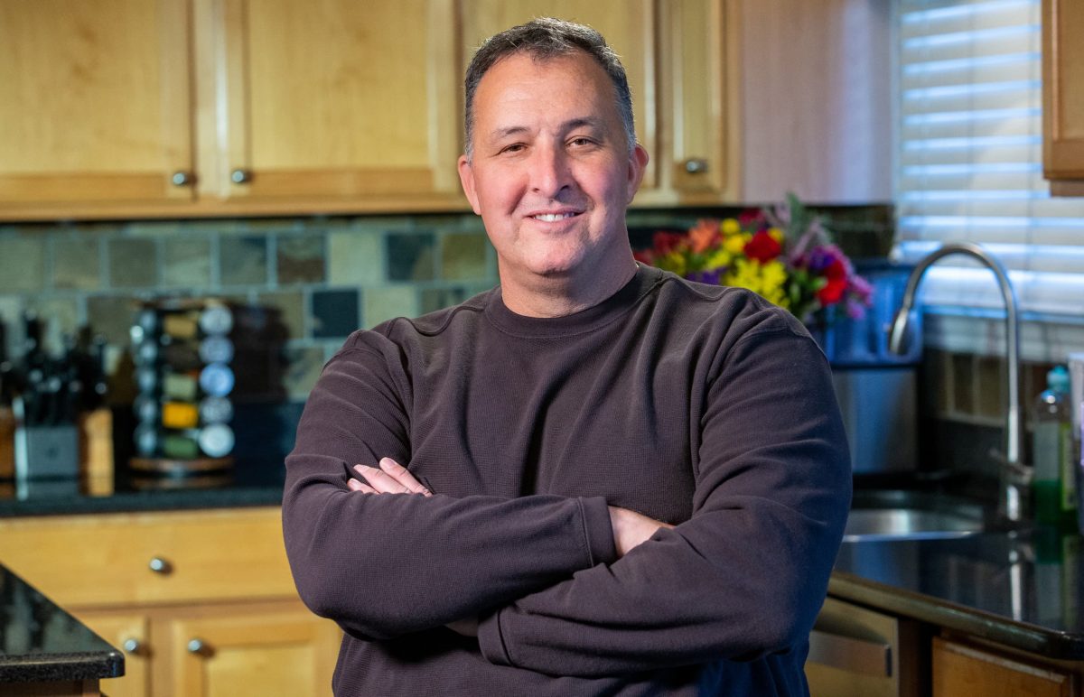
Like any surgery, there can be side effects, like in Valerie’s case. Because of the location of her tumor, she lost her ability to smell. For Stephen, he experienced added restrictions as he waited for his vision to return to normal once the tumor was removed. Kevin didn’t experience difficulties after his surgery, but he did work hard to regain his strength and balance.

If you or a loved one is diagnosed with a brain tumor, consider all of your options and seek a second opinion. Like Valerie, you might find an option you didn’t expect.


Watch Stephen’s brain tumor story below:

Watch how a large pituitary tumor interrupted Kevin’s life:

Watch Valerie’s full story below:


To schedule an appointment with Dr. Jacob or any of the neurosurgeons at MHSI, call 248-784-3667 or visit MHSI.us

race47361-social1200x630.bzcJmm.jpg

September marks another effort to raise awareness about Chiari malformation. Chiari malformation is a disease of the brain where brain tissue is pushed down into the spinal canal. It occurs when the base of the skull is abnormally small or misshapen, causing pressure on the brain which forces it downward. Chiari malformation may cause blockage in the flow of the cerebrospinal fluid, which may cause the normal fluid spaces in the brain to enlarge, also known as hydrocephalus.

It was estimated that the condition occurs in about one in every 1,000 births. However, the increased use of diagnostic imaging has shown that Chiari malformation may be much more common. Complicating this estimation is the fact that some children who are born with the condition may not show symptoms until adolescence or adulthood, if ever. It can also be hereditary, and often those with Chiari will recall a family member with the same symptoms. Family members such as siblings Ezra and Katelyn, and mother Tammy and daughter Sheena.

Watch Ezra and Katelyn’s journey with Chiari below:

Chiari Malformation Corrected for Mother Tina and Daughter Sheena:

Adults and children alike can go through life and from doctor to doctor, with severe symptoms and are not diagnosed until an aware physician recognizes the symptoms. Patients Tina, Lu Anna and Tyler each had a physician acknowledge their symptoms and refer them to the leading expert in Chiari decompression, Neurosurgeon Dr. Holly Gilmer.

To learn more about Chiari malformation, the symptoms, and treatments, click here.

For most patients, the diagnosis is elusive for many years until the patient meets a doctor who is aware of Chiari, and understands the symptoms presented can become more manageable with surgery. One of the goals of Conquer Chiari Walk Across America is to raise awareness and funds to support further research. At this event, patients, their families and friends, along with doctors join in to walk to raise funds every third and fourth Saturday in September.

Click the graphic below to download the flyer:

Conquer Chiari Walk Across America


If you or a loved one is experiencing symptoms of Chiari malformation, call 248-784-3667 or visit MHSI.us to schedule an appointment with Dr. Gilmer.


BrainTumor-1250205787-1200x772.jpg

Medical illustration of brain tumor

It doesn’t get much more serious than being given the diagnosis that you have a brain tumor. Though brain cancer is rare and may be a life-threatening situation when detected, many forms of tumors can be successfully treated.

A brain tumor is a collection, or mass, of abnormal cells growing in, or next to the brain. But, what are the different types of brain tumors, and what are the symptoms to look out for?

Brain Tumor Symptoms
There are so many different types of brain tumors, and because each is so complex, brain tumors can cause different effects for each person. Physical problems and behavioral changes can occur throughout the body severely impacting one's life. The symptoms will depend on the individual's particular tumor, its location, and can result in:

  • Drowsiness
  • Personality changes
  • Confusion
  • Impulsiveness
  • Blurred vision
  • Balance problems
  • Headaches
  • Nausea
  • Vomiting
  • Seizures

Types of Treatable Brain Tumors
Though there are many different types, a brain tumor is classified by where it was originally formed. If it originated in the brain, it’s called a primary tumor and can be either noncancerous (benign), or cancerous (malignant). A tumor that originates in another part of the body and travels to the brain is called a meta tumor, and they are always cancerous.

Metastatic Brain Tumors - Also known as secondary tumors, they originate outside of the brain in another part of the body and then spread to the brain.

Meningiomas - Originating in the meninges, these thin layers of protective tissues surround the brain and spinal cord. Meningiomas usually grow slowly and most are not cancerous.

Pituitary Tumors - Found just under the brain, the pea-sized pituitary gland makes hormones that affect many of the body’s functions. A pituitary tumor can cause it to release too much, or too little, of these hormones which can cause serious problems.

Glioblastoma Multiforme - These are the most challenging forms of brain tumors to treat. MHSI physicians have access to the most current technology to deal with this cancer including image-guided surgical treatments.

Schwannoma - The most common type of benign peripheral nerve tumor in adults and rarely cancerous. When schwannoma grows larger, it can make removal more difficult. This type of nerve tumor can occur in any part of the body, and at any age.

Keith’s Brain Tumor Story
Before Keith, a patient at Michigan Head and Spine Institute, was diagnosed with his brain tumor he was an accomplished musician hoping for a big break.

“I got my first guitar when I was 12. It’s what calms me. It’s where I can focus on one thing and the thousand thoughts going on in my head will go away.”

But, that was all about to change when he started experiencing unexplained health problems and physical ailments.

“The first symptoms I had were the nausea, dizziness and headaches,” he said, “It got to the point where I couldn’t eat. The only thing that was staying down was water.”

Knowing something was wrong with his health, he went to an emergency room hoping it was just an ear infection. After undergoing some tests, his ER doctor came back with unsettling information.

“A brain tumor was not on my list.” - Keith, a patient at MHSI

Diagnosed with a sporadic case of hemangioblastoma, a benign tumor, Keith was seen by Michigan Head and Spine Institute, trusted experts for treatment and diagnosis of tumors, diseases and other conditions of the brain.

After reviewing his CT scan, it was confirmed Keith had a large mass at the back part of the head in the region of the cerebellum. This part of the brain also contributes to coordination.

“I just wanted it to stop hurting, to stop feeling sick,” Keith recalled.

While being prepped for surgery he also remembered the last thing he told the anesthesiologist, “I said, ‘I’m a musician.’ When I came back out, I still wanted to be a musician.”

Keith’s surgery was successful. He then underwent a second nonsurgical procedure called Gamma knife treatment to remove the last bit of his tumor.

“When I woke up after the surgery, I knew it was fixed. The pain I was having before was gone,” he said relieved, “I like the way my doctor had a very confident way about himself.”

As for Keith’s guitar playing and music career after his surgery at MHSI:

“I’ve been in a few bands, had a little stardom,” he updated, “I’ve met a lot of people in the industry since the surgery that say they’re going to help me, so we’ll see what happens.”

To learn more about conditions of the brain and brain tumor procedures, please visit our MHSI Patient Education page at: Conditions of the Brain.

If you or a loved one is seeking treatment for a condition of the brain, or are seeking a second opinion on an existing diagnosis, schedule an appointment online or contact MHSI at 248-784-3667.

To view more information about MHSI’s neurosurgeons, visit: https://www.mhsi.com/doctors/neurosurgery-physicians/. For more information about MHSI’s  neurologists, visit: https://www.mhsi.com/doctors/neurologists/


DBS_Thumbnanail.jpg

Movement disorders refer to conditions which cause abnormal voluntary or involuntary movements. Symptoms are caused by disorganized electrical signals in specific areas of the brain. Those challenged with essential tremor, Parkinson’s disease, dystonia, and other neurological conditions may benefit from deep brain stimulation (DBS), especially when symptoms progress or medications become less effective.

DBS is performed by neurosurgeons who precisely place one or more wires, called electrodes or leads, inside the brain. The lead is connected to a pacemaker-like device called a neurostimulator that is surgically implanted into the patient’s chest. The neurostimulator then conducts continuous pulses of electric current through the leads to interrupt the disorganized brain signals causing tremor or other movement disorder symptoms.

Insertion of the leads and neurostimulator are typically accomplished in two steps. First, the neurosurgeon places the leads either into one or both sides of the brain, and this is often performed while the patient is awake. In a second procedure the neurostimulator is implanted in the chest while the patient is asleep. Programming of the neurostimulator can be performed by the neurosurgeon, neurologist, or primary care physician, to find the optimum settings that are effective for each patient. There are different devices available that can tailor therapy to each individual patient, and the device representative often has an important role to help the patient become familiar with their programmer and programming settings.

For patients with essential tremor, the most common movement disorder, DBS can return a person to normal daily activities like dressing, shaving, eating, and drinking. Symptoms of Parkinson’s disease such as tremor, bradykinesia and rigidity are especially well-treated with DBS, and medication dosages can often be decreased. Dystonia is an uncommon movement disorder, with symptoms of abnormal posturing and twisting movements, which also respond to DBS.

DBS can also be used to treat patients with epilepsy, obsessive-compulsive disorder, or certain pain disorders. These unique disorders are often managed by specialized neurologists and psychiatrists, and a thorough discussion with your specialist is necessary to determine if you are a candidate for this therapy.

At MHSI, a neurosurgeon or functional neurosurgeon will work with a patient’s neurologist or primary care physician prior to and after surgery to ensure that the optimal therapy is achieved.

For more information about DBS, visit our patient education page on the topic, click here. If you or a family member or friend might benefit from DBS, please schedule a consultation by calling 248-784-3667 or visit MHSI.us.

MHSI Neurosurgeons Who Perform DBS 

Dr. Fredrick Junn
Dr. Richard Veyna


Zeke_D110515-2252.jpg

Every year, more than 200,000 people are diagnosed with Chiari Malformation, a condition where a portion of the brain pushes into the spinal column, reducing the flow of spinal fluid.

Receiving a Chiari diagnosis is often a relief just to know that the symptoms are acknowledged.

Chiari Malformation can cause severe headaches, dizziness, difficulty swallowing and more. Adults have balance issues, debilitating headaches, muscle weakness, ringing in the ears and neck pain. Infants and children experience developmental delays, gagging, vomiting or reflux, excessive drooling and irritability.

This combination of symptoms can make life miserable. Fortunately, MHSI has one of the nation’s foremost experts in the surgical treatment of Chiari Malformation, Holly Gilmer, M.D., who has the knowledge and experience in offering treatment to both adults and children. Patients have traveled as far as Colorado, Maine, and Arizona to Michigan just so they could have the best chance at a full recovery.

Amy, a kindergarten teacher, is a patient of Dr. Gilmer. Battling extreme exhaustion and migraine headaches, Amy was diagnosed with Chiari Malformation, but the first neurosurgeon she saw told her the treatment is an elective surgery. When her daughter was diagnosed with a tethered spine, Amy found Dr. Gilmer.

“I kept going to doctors and they kept telling me I was stressed out and overworked. They gave me anxiety pills,” recalled Amy.

After meeting with Dr. Gilmer and choosing Chiari decompression surgery, Amy is off her headache medications, no longer has a stiff neck and, in general, feels great.

Patients who are diagnosed with Chiari Malformation and choose neurosurgery at Michigan Head and Spine Institute have comfort in the knowledge that Dr. Gilmer literally wrote the book on Chiari Malformation and decompression surgery. Her work is referenced in medical journals and with Chiari patients all over the world.

In addition to Dr. Gilmer’s expertise, patients at MHSI are in excellent hands throughout their care, no matter what their diagnosis. Throughout the spectrum of your treatment – from making your first appointment, through diagnosis and on to surgery and recovery – every patient deserves the best. Our experience and expertise gives MHSI patients an advantage for an excellent recovery.

Watch Amy’s and Zeke’s stories in their own words


karol-thumb.jpg

<div class="at-above-post-cat-page addthis_tool" data-url="https://www.mhsi.com/blog/minimally-invasive-spine-surgery/"></div>The physical symptoms of spine and neck conditions can be debilitating – and the mental side effects can be worse.<!-- AddThis Advanced Settings above via filter on get_the_excerpt --><!-- AddThis Advanced Settings below via filter on get_the_excerpt --><!-- AddThis Advanced Settings generic via filter on get_the_excerpt --><!-- AddThis Share Buttons above via filter on get_the_excerpt --><!-- AddThis Share Buttons below via filter on get_the_excerpt --><div class="at-below-post-cat-page addthis_tool" data-url="https://www.mhsi.com/blog/minimally-invasive-spine-surgery/"></div><!-- AddThis Share Buttons generic via filter on get_the_excerpt -->


TrentonOfficeGraphic.jpg

Downriver Communities Now Have Neurosurgery Expertise

When looking for a neurosurgeon, you want someone you can trust with head, neck and spine conditions. Someone who has expertise with a good reputation. Someone close to home.

MHSI opened a new office, conveniently located on the campus of Beaumont Hospital, Trenton. Staffed by neurosurgeons Ratnesh Mehra, D.O., Pradeep Setty, D.O.  and Jeffrey Jacob, M.D., we offer the caliber of care and treatment you expect, only closer to home.

Neurosurgery is delicate, and you want the best, most experienced surgeons. The team at MHSI – Trenton has a complex set of skills including minimally invasive and robotic surgery, superior knowledge of brain tumors, and leading-edge experience with skull base and spine surgery.

Patients have long traveled to MHSI to be seen by specialists who provide the full spectrum of care for head, neck and spine conditions, because we provide a range of treatment and diagnosis options that are unparalleled in Southeast Michigan. And with the new Trenton office so close to home, receiving our care just got easier.

This is our tenth location and our first Downriver. Opening another office cements our mission to you: Provide the best, most experienced care. Spine, neck and head conditions are complex, and you want the best surgeon. With the team at MHSI working for you, helping you make the best decisions for your health, you know you have the professionals you want on your side.

The new location on Fort Street, on the campus of Beaumont Hospital, Trenton, opens up conveniences for patients as well. Imaging, testing, therapies and more are within arm’s length at the hospital, while the doctors you trust are right there with you.

We are accepting new patients, seeing post-op patients and continuing to provide care for those in the follow-up stage of care. You can call 248-784-3667 for an appointment, or request one online.

Expertise makes a difference, and the difference is MHSI.