Spine Archives - Michigan Head & Spine Institute Blog

Steele-Xray-Blog_Scoliosis-945206642_MHSI_blog.jpg

Dr. John J. Steele

John J. Steele, M.D., Ph.D.
Neurosurgeon

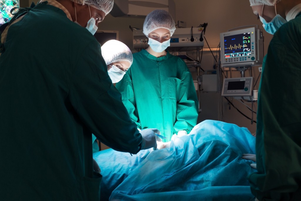
You may not be familiar with the O-arm™, but this imaging system has revolutionized spinal surgery, along with other surgical procedures. Getting to know the O-arm™, including what makes it superior to other imaging tools and why it’s considered top-of-the line for spinal trauma surgeries, can help you understand why Michigan Head & Spine Institute surgeons sing the praises of this system.

What is the O-arm™?

The O-arm™ system is a mobile X-ray system that provides 2D and 3D images during surgical procedures. It provides visibility that allows surgeons to be ultra-precise, helping them pinpoint problems and take care of them with minimal impact on the patient. It also helps surgeons apply medical devices in a way that uses the smallest and fewest of those devices — yet provides the strongest possible repair. The O-arm™ also exposes patients to less radiation than other surgical imaging tools.

How Does the O-arm™ Help Spinal Surgery Patients?

Surgery is all about precision. Thanks to the imaging provided by the O-arm™, surgeons can be more precise than ever, which often means fewer or smaller incisions and faster recovery time for patients.

The O-arm™ also affords surgeons the ability to precisely navigate complex surgical situations in a way that wasn’t previously possible. Consider, for example, a congenital spinal problem such as a sacral meningocele. To operate in such a “busy” area near the spine, surgeons need to be confident in their ability to be precise. The O-arm™ affords them this confidence.

Another cutting-edge application of the O-arm™ involves the placement of cervical pedicle screws. The pedicle is like the yellow brick road of the spinal column, and pedicle screws help support the weakest part of the spine. The computer precision of the O-arm™ allows surgeons to place pedicle screws accurately and efficiently in a much less invasive way than other, previously used imaging tools. This type of surgery wasn’t possible 10 years ago, or even two years ago.

Yet another major benefit of the O-arm™ is that it allows surgeons to check in real time that they’ve placed devices correctly or take another image after removing something during surgery. If they see something additional that needs to be corrected or removed, they can take care of it while the patient is still under anesthesia, without subjecting them to another procedure.

Other Applications of the O-arm™

As the O-arm™ system becomes more widely available, it is being explored as a tool for many different kinds of procedures. In addition to spinal trauma surgery and procedures to address congenital spine problems, it is also being used to help patients with scoliosis.

Outside of the spine, the O-arm™ is being considered for other types of trauma and orthopedic surgery. As the number of uses for this tool grow, it’s even possible that in the next 10 years the O-arm™ will be used to place deep brain stimulators because it provides such an accurate roadmap.

Much like NASA’s Artemis program is helping us explore space like never before, the O-arm™ is catapulting us into a new neurosurgical universe.

If you or a loved one is seeking treatment for neurosurgery, or to schedule an appointment with Dr. Steele or any of our MHSI experts, visit www.mhsi.com/appointments or call 248-784-3667.


Brain-Surgery-1306095257_MHSI-1200x683.jpg

Although it’s relatively new, endovascular neurosurgery is being used to treat an increasing number of health problems — with remarkable results. Through research and collaboration, experts from a variety of fields are coming together to innovate a type of surgery that is helping some of the highest risk patients.

What is Endovascular Neurosurgery? 

Endovascular neurosurgery is a subspecialty of neurosurgery that combines multiple types of expertise. Incorporating both neurosurgery, which treats conditions of the brain and nervous system, and interventional radiology, which uses imaging and minimally invasive techniques to diagnose and treat injuries and health conditions, endovascular neurosurgery uses small catheters to diagnose and treat health problems affecting the brain and spinal cord.

What Conditions Endovascular Neurosurgery Treat?

Endovascular neurosurgery can be used to diagnose and treat a variety of injuries, diseases and health conditions including:

  • Strokes
  • Brain aneurysm
  • Cerebrovascular malformations including arteriovenous malformations (AVMs) and dural arteriovenous fistula (dAVF)
  • Carotid artery disease
  • Atherosclerosis (narrowing) of the vessels of the brain
  • Idiopathic intracranial hypertension (formerly pseudotumor cerebri)

This is merely a partial list of the many health concerns that can be addressed with endovascular neurosurgery — a list that will continue to grow as neurosurgeons, interventional radiologists, and interventional neurologists work together to advance the science surrounding these techniques.

What Are the Benefits of Endovascular Neurosurgery?

The blood vessels of the brain are deep within the brain itself, meaning they’re not readily accessible without open surgery. Because it involves the use of catheters to pass through blood vessels, endovascular neurosurgery is a minimally invasive technique that provides an alternative to more invasive options.

Rather than opening the skull to get into the brain, endovascular neurosurgeons often only need to make a tiny puncture to insert the catheters they use in surgery.

The minimally invasive nature of endovascular neurosurgery means that some patients who receive elective (non-emergency) types of this procedure can go home the same day.

Even in cases where recovery takes a little longer, patient outcomes are generally very good. Many people can go home within one day of surgery and are back at work within a week.

Another major benefit of endovascular neurosurgery is that it offers hope for people with diseases or conditions that would have been otherwise untreatable. These are patients who didn't have a different treatment option — or at least a good treatment option — and these endovascular techniques are allowing neurosurgeons to provide life-altering or even life-saving care for them.

Advancing Technology, Saving Lives 

Endovascular neurosurgery is an exciting field that allows surgeons to greatly improve outcomes for patients who were otherwise at the mercy of their disease.

Treatment options that weren’t available only a few decades ago are now saving people’s lives — and will continue to do so as the field grows and expands through continued collaborative research and innovation.

If you or a loved one is seeking treatment for a condition of the spine, or to schedule an appointment with any of our MHSI experts, visit www.mhsi.com/appointments or call 248-784-3667.


karol-thumb.jpg

<div class="at-above-post-cat-page addthis_tool" data-url="https://www.mhsi.com/blog/minimally-invasive-spine-surgery/"></div>The physical symptoms of spine and neck conditions can be debilitating – and the mental side effects can be worse.<!-- AddThis Advanced Settings above via filter on get_the_excerpt --><!-- AddThis Advanced Settings below via filter on get_the_excerpt --><!-- AddThis Advanced Settings generic via filter on get_the_excerpt --><!-- AddThis Share Buttons above via filter on get_the_excerpt --><!-- AddThis Share Buttons below via filter on get_the_excerpt --><div class="at-below-post-cat-page addthis_tool" data-url="https://www.mhsi.com/blog/minimally-invasive-spine-surgery/"></div><!-- AddThis Share Buttons generic via filter on get_the_excerpt -->


Research-1024x1024.jpg

Over two-thirds of individuals experience chronic low back pain (LBP) in their lifetime, according to the New England Journal of Medicine. Some of the most common causes of low back pain include the biochemical degeneration of the intervertebral disc (IVD), spinal stenosis, and disc herniation. For these individuals, posterolateral and interbody fusion techniques are frequently considered for those with one- or two-level degenerative disc disease whose symptoms are unresponsive to conservative treatment. Nevertheless, pre-operative diagnostic techniques that may identify those patients with degenerative disc disease without significant neurological compression who may benefit from surgical intervention remain elusive.

Henry C. Tong, M.D. of MHSI and his colleague at Oakland University William Beaumont School of Medicine, Mengqiao Alan Xi, BSc, recently published their research that re-evaluated the effectiveness of lumbar discography with post-discography CT.

Their study results indicate that discography with post-discography CT can be an effective method to evaluate patients with discogenic back pain refractory to non-operative treatments. Those patients with one- or two-level high concordant pain scores with associated annular tears and negative control disc represent good surgical candidates for lumbar interbody spinal fusion.

To read the full study, click this link.


surgery-dreamstime_xl_35011296-1024x683.jpg

Jeffrey Jacob, M.D.
Jeffrey Jacob, M.D.

Jeffery Jacob, M.D., along with his former Mayo Clinic colleagues in the Department of Neurosurgery and Anesthesia.  The article was published in Neurology, Sept. 8, 2015, which presented findings from a study on obese patients undergoing elective spinal fusion.

The objective of the study was on the impact of obesity on resource utilization and early complications in patients undergoing surgery for degenerative spine disease.

The conclusions drawn from this single-institution retrospective analysis is that increased BMI is associated with longer operative times, increased complication rates and increased cost independent of co-morbidities. The results further indicated the need for preoperative weight loss.